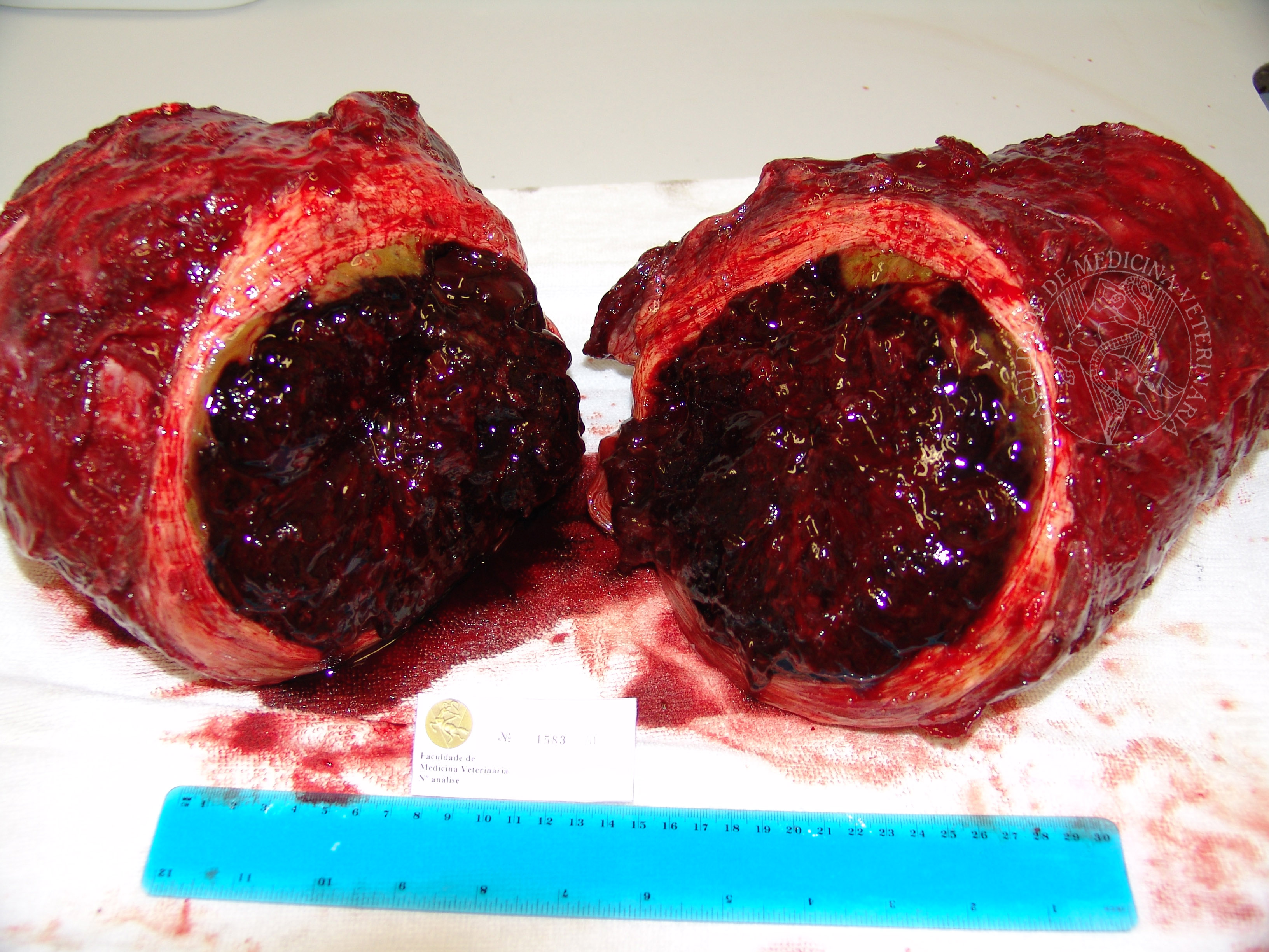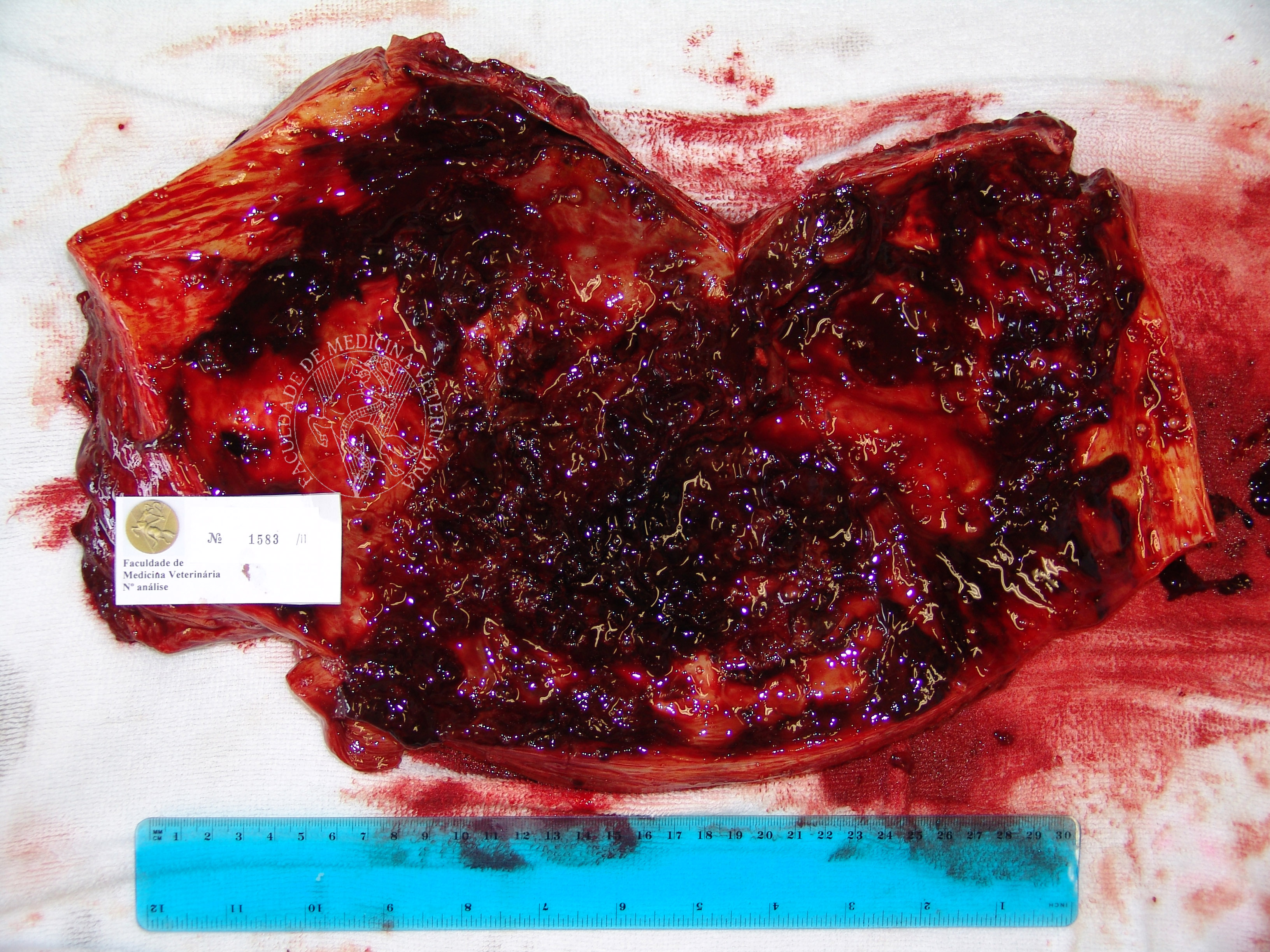

Telangiectasic osteosarcoma in a horse. This subcutaneous mass, located over the right iliac tuberosity, developed over several months, finally reaching its final size (50 by 30cm, 8Kg) after the animal was taken out of the field for training. Its thick wall, made of mesenchymal neoplastic cells arranged in multidirectional beams of acidophil material, encased an exuberant blood clot. Once removed, the mass’ inner lining was revealed to be smooth and regular. The presence of several areas of well differentiated bone tissue, as well as the strong hematic component of the mass, determined a final diagnosis of osteosarcoma, telangiectasic variant.
 |
 |
 |
Portuguese
English
 |
 |
 |
Loading
Copyright © 2001 by MC Peleteiro. M Pinho & JS Orvalho
design by R Noiva